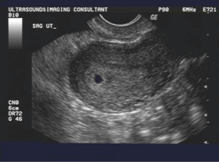
Normal first trimester ultrasound - Normal first trimester 6 weeks ultrasound.


The connections between the lateral ventricles and third ventricle foramina of Monro are still wide.

These findings suggest that the phenotypic expression of the syndrome is evident from at least 11 weeks of gestation.
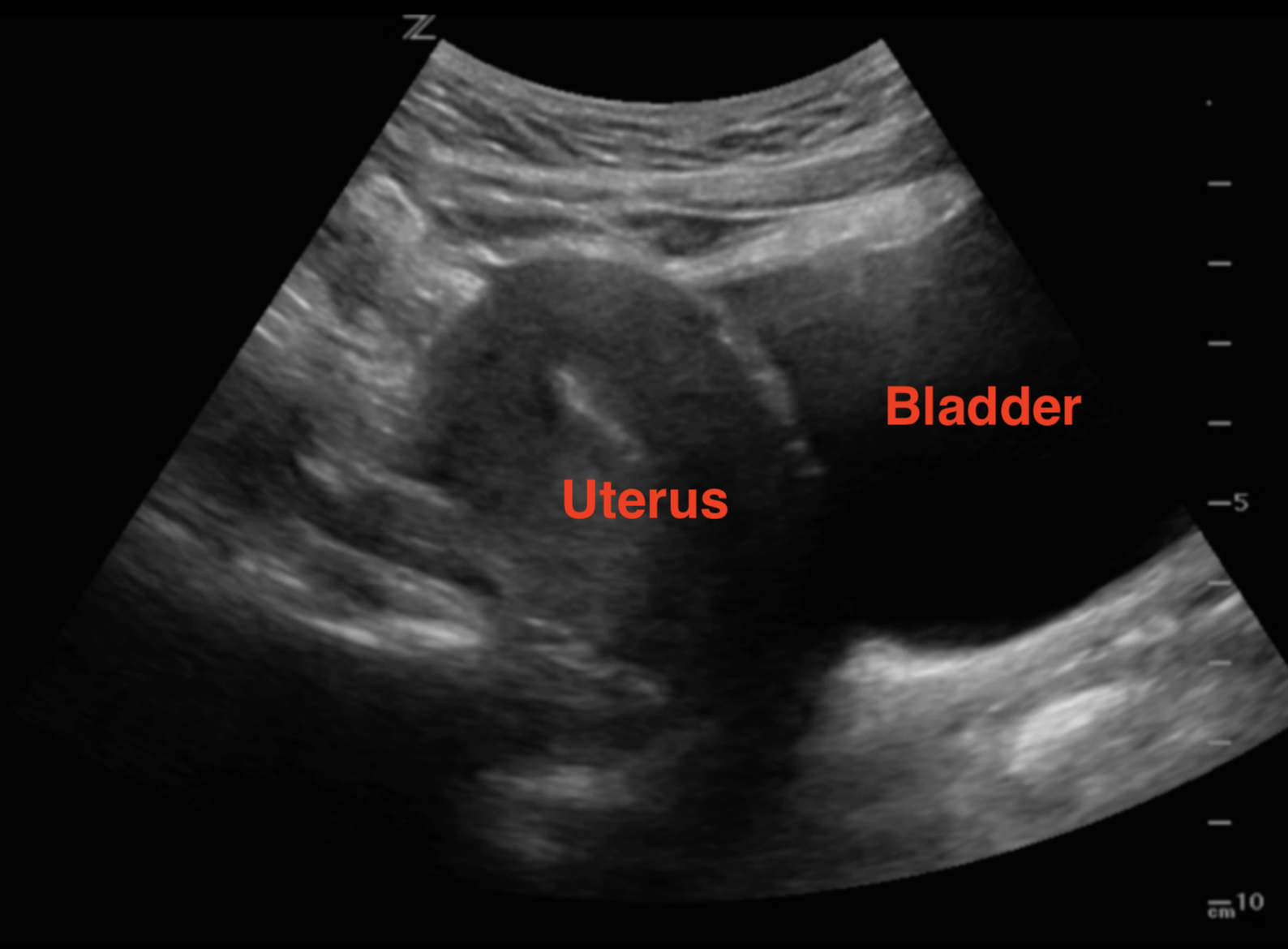
The diameter of the yolk sac increases between 5 to 10 weeks to a maximum of about 6mm, and the measurement of the yolk sac is usually from inner wall to inner wall.


Fetal ultrasound
OB Images
Both types of scans typically last about 20 minutes and are painless.
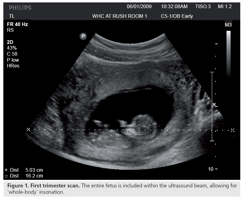
The diagnostic possibilities in this setting are a normal intrauterine pregnancy that is too early to be visualized, a failed intrauterine pregnancy miscarriage , and an ectopic pregnancy.
Description: By week 8, it is regularly identified.
Sexy:
Funny:
Views: 1526
Date: 12.02.2022
Favorited: 119

Category: DEFAULT
User Comments 3

Chorionic villus sampling showed Turner mosaicism and the pregnancy was terminated.
More Photos
Latest Photos
Latest Comments
- +127reps
- Speakers will disclose any relevant commercial relationships prior to the start of the educational activity.
- By: Grote
- +955reps
- Sonographers can be trained to perform intracranial translucency measurements at the 11 to 13 week exam, resulting in reproducible results.
- By: Gewirtz
- +772reps
- For the ultrasound studies, 7.
- By: Burtis
- +517reps
- The double sac sign refers to the presence of two concentric echogenic rings surrounding at least part of the fluid collection, hypothesized to represent the decidua capsularis and parietalis.
- By: Honniball
- +54reps
- If viability remains unclear, repeat transvaginal ultrasound in 11 to 14 days.
- By: Pistol
4cq.net - 2022
DISCLAIMER: All models on 4cq.net adult site are 18 years or older. 4cq.net has a zero-tolerance policy against ILLEGAL pornography. All galleries and links are provided by 3rd parties. We have no control over the content of these pages. We take no responsibility for the content on any website which we link to, please use your own discretion while surfing the porn links.
Contact us | Privacy Policy | 18 USC 2257 | DMCA


